
Faculty
Daniela Bacherini
Country: Italy
Affiliation: Eye Clinic, University of Florence, AOU Careggi
Bio:
Daniela Bacherini graduated in Medicine and Surgery in 2008 with full marks from the University of Florence, discussing a thesis in ophthalmology in the field of medical retina.
Webinar


29 March 2022
WATCHDiabetic retinopathy: clinical patterns and management of macula and peripheral retina.


June 17, 2026, 6.30 PM CEST
WATCHFrom intermediate AMD to geographic atrophy: toward a personalized therapeutic approach

Videos

Image Bank
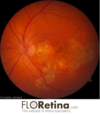
TUBERCULOUS SERPIGINOUS-LIKE CHOROIDITIS 1
Daniela Bacherini Lorenzo Vannozzi45 Y/O female with serpiginoid lesion extending from the juxtapapillary area. DEVICE: Fundus Camera, Zeiss
View image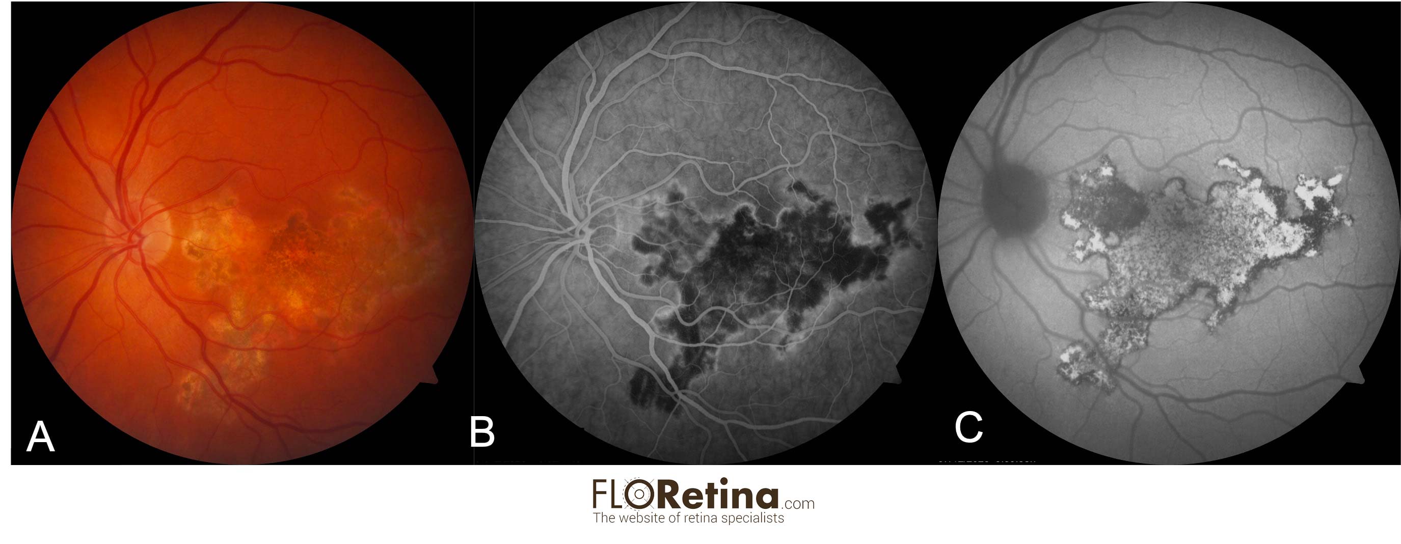
TUBERCULOUS SERPIGINOUS-LIKE CHOROIDITIS 2
Daniela Bacherini Lorenzo VannozziColor fundus photograph (A) 45 Y/O female with serpiginoid lesion extending from the juxtapapillary area. Intermediate-phase fluorescein angiogram photographs (B) of the same eyes delineating the typical hyperfluorescent margins of the serpiginoid lesions. Fundus autofluorescence image (C) of the same eye, disclosing predominantly hyperautofluorescent lesion with stippled hypoautofluorescence delimited by a thin rim of hypoautofluorescence. DEVICE: Multimodal imaging: (A) Color fundus photograph (Zeiss), (B) FA (Heidelberg), (C) Fundus autofluorescence (Heidelberg).
View image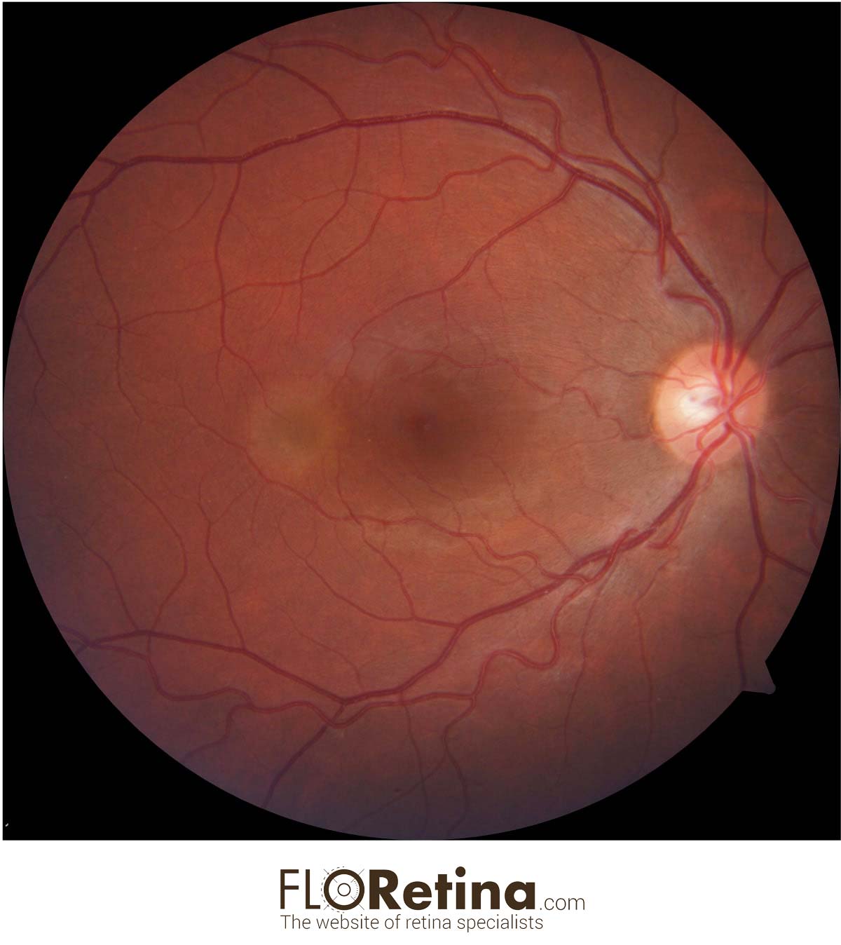
UNILATERAL ACUTE MACULOPATHY CAUSED BY COXSACKIEVIRUS
Daniela Bacherini Stefano Mercuri Federica Serino37 Y/O male with an eccentric yellow lesion (orange arrow) corresponding to a neurosensory retinal detachment. DEVICE: Fundus camera, Zeiss.
View image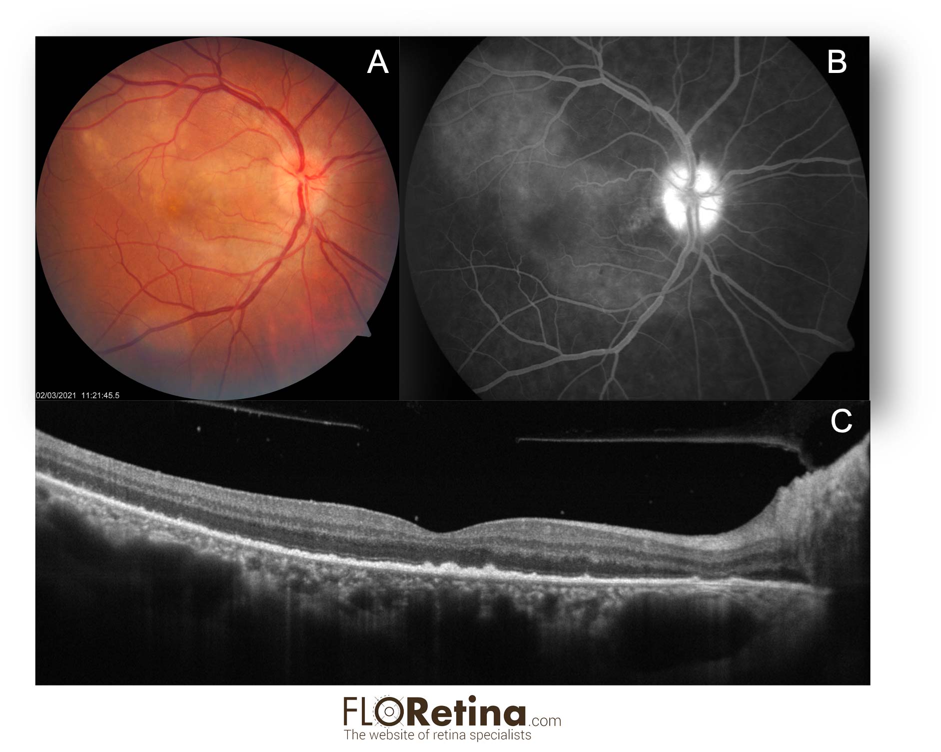
Acute Syphilitic Posterior Placoid Chorioretinopathy (ASPPC)
Daniela Bacherini Stefano Mercuri Fabrizio GiansantiA. Fundus photograph showing vast round placoid yellow zone in the posterior pole;
B. FA reveals hyperfluorescence of the optic disk with a zone of hyperfluorescence in the posterior pole (tissue staining).
C. Structural OCT shows disruption of the ellipsoid zone and hyperreflective, nodular thickening of the RPE. Hyperreflective dots representing inflammatory cells are visible in the vitreous and attached to the posterior hyaloid.
DEVICE: Multimodal imaging: fundus photograph, FA, Structural OCT
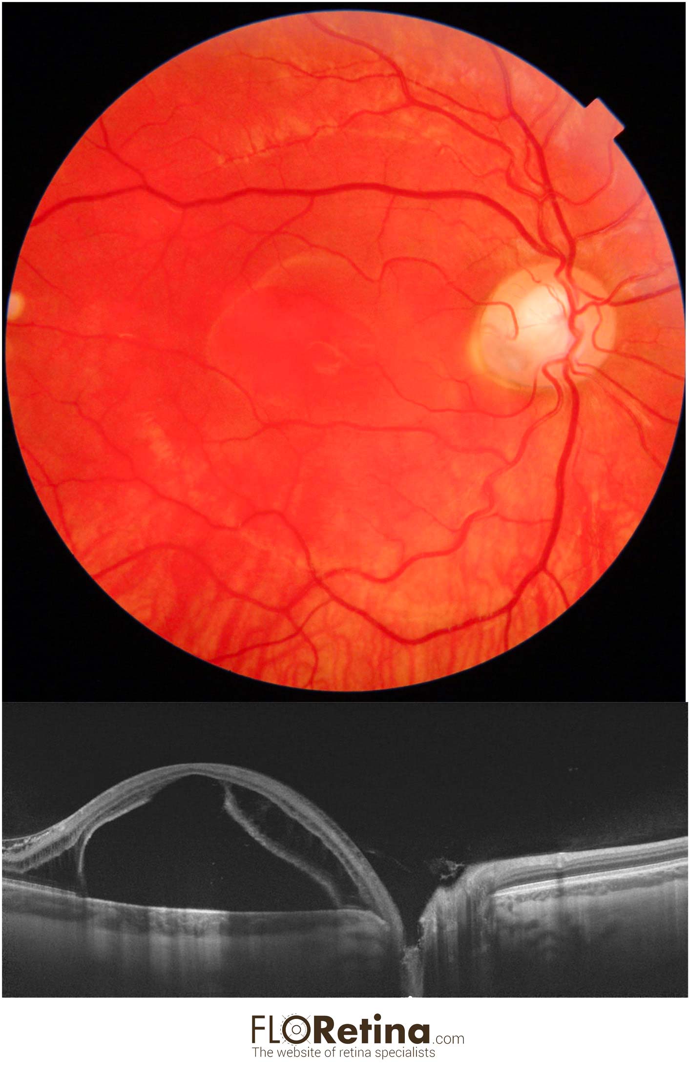
OPTIC DISK PIT
Daniela Bacherini Tomaso Caporossi Alfonso Savastano25 y/o male with visual reduction showing macular schisi with detachment due to optic coloboma.
A. Color fundus photograph
B. Structural OCT showing the retina splitted at both the inner and outer retinal layers with a macular detachment
DEVICE: Fundus photograph (Topcon), Structural OCT (Topcon)
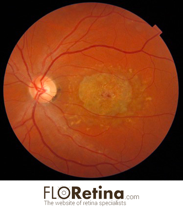
STARGARDT DISEASE
Daniela Bacherini34 yo female with atrophic maculopathy and adjacent flecks. DEVICE: Color fundus photograph (Tocpon)
View image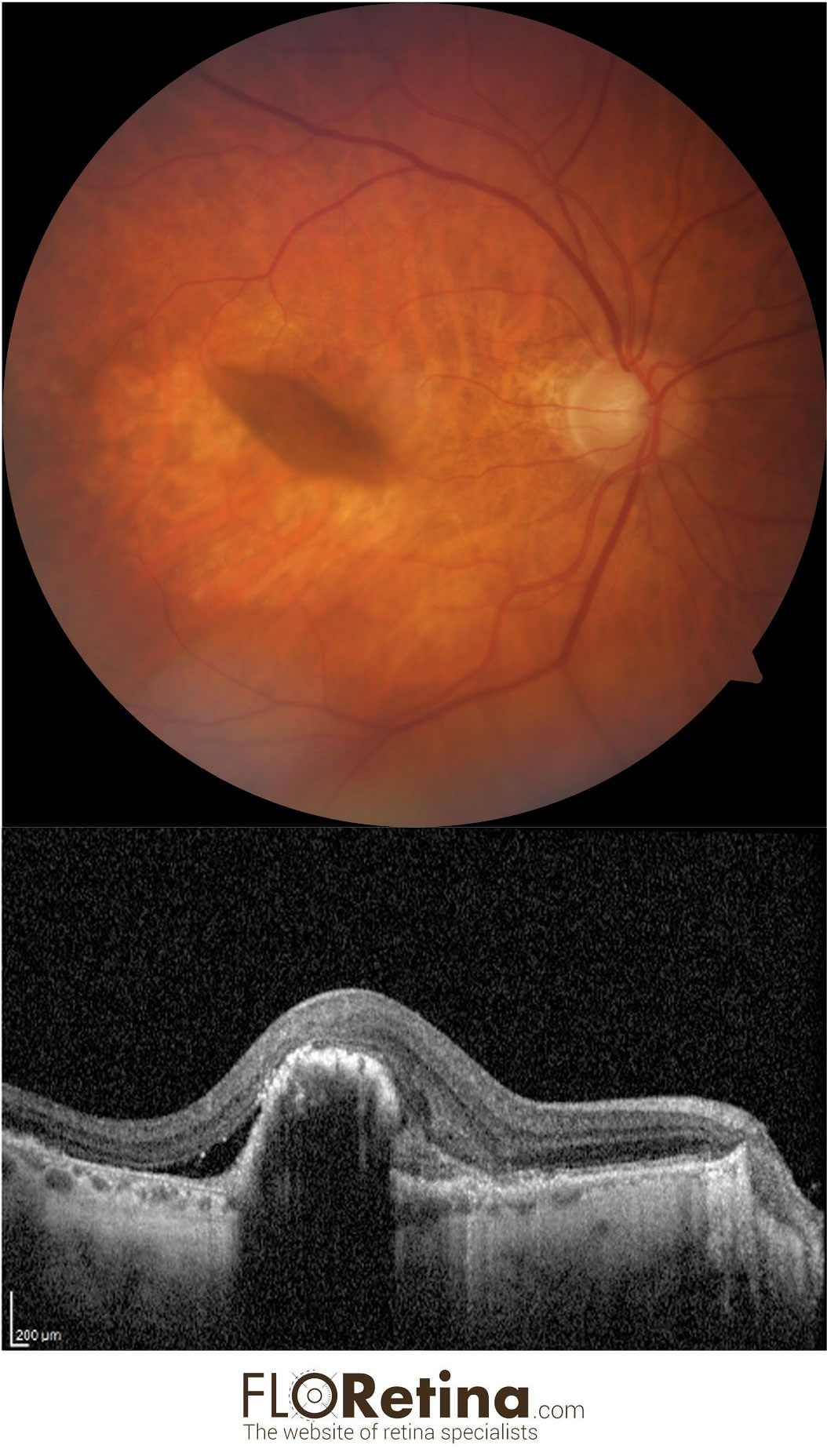
RPE (retinal pigment epithelium) tear
Daniela Bacherini78 yo female with RPE tear. DEVICE: Fundus fotograph (Zeiss), structural OCT (Heidelberg)
View image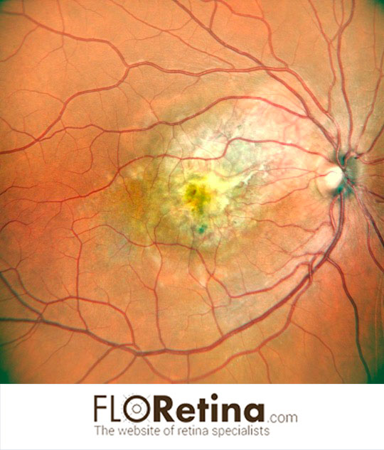
CHOROIDAL OSTEOMA complicated by choroidal neovascularization
Daniela Bacherini Stefano Mercuri Fabrizio GiansantiImaging device: Color fundus photography, angle 89°, Nidek
View image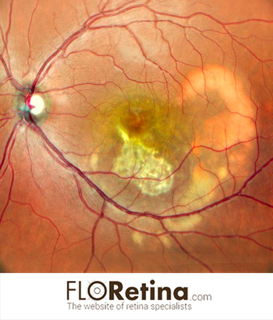
Extramacular Best disease
Daniela Bacherini Francesca SantoroImaging device: Color fundus photography, angle 89°, Nidek
View image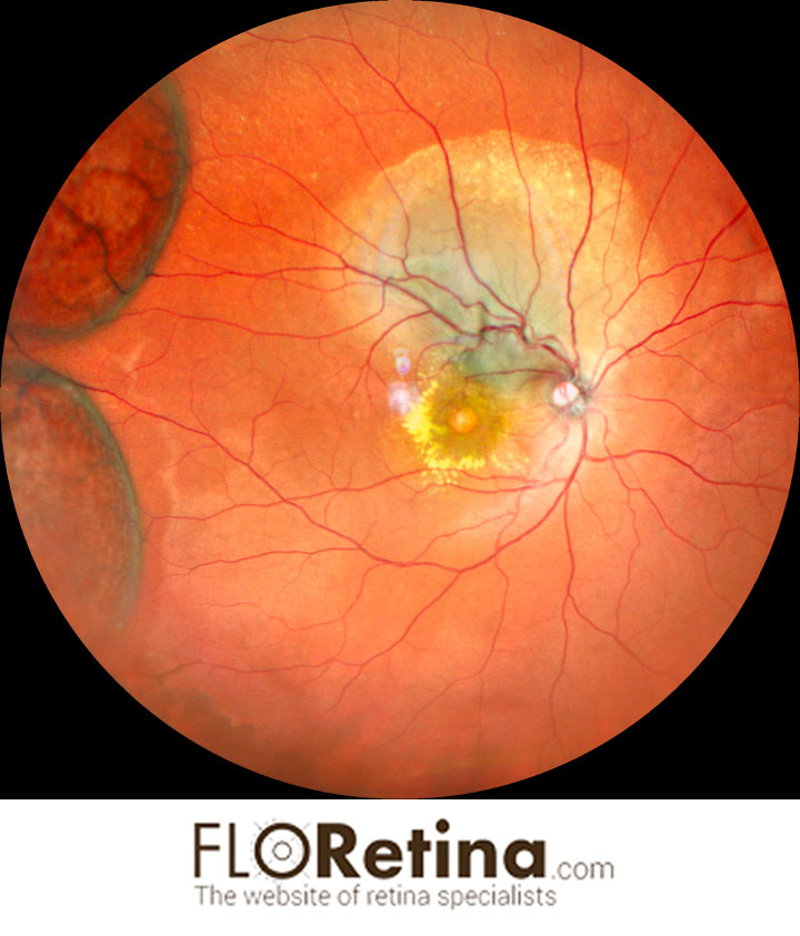
Choroidal tubercoloma and peripheral retinoschisis
Daniela BacheriniImaging device: Ultrawidefield color fundus photograph 163°, Nidek
View image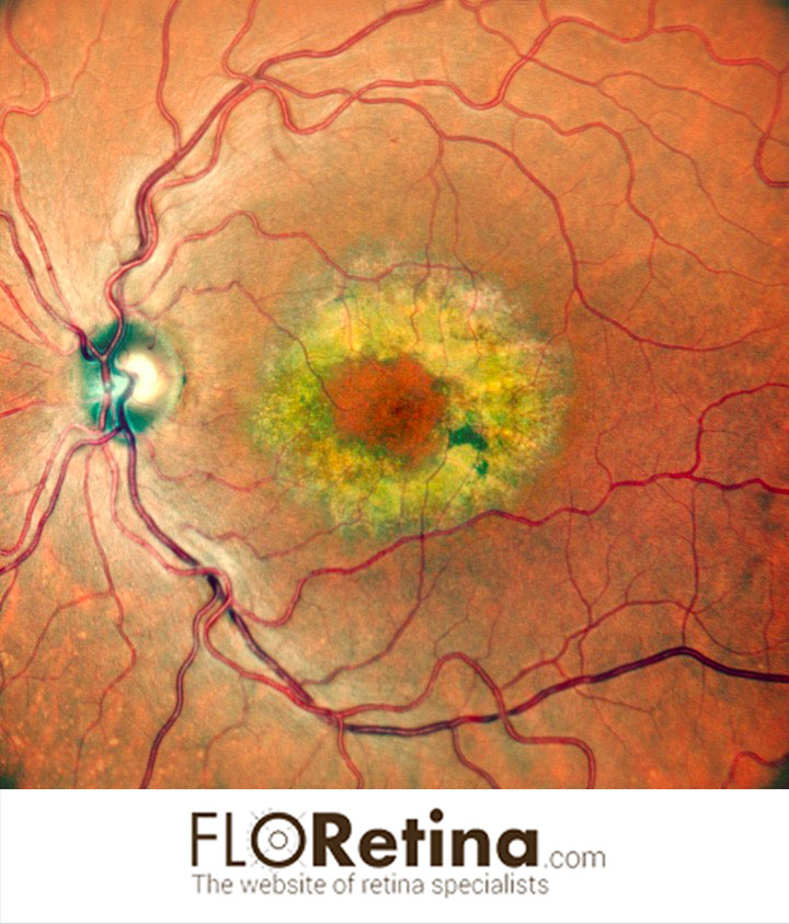
Benign Concentric Annular Macular Dystrophy
Daniela BacheriniImaging device: Color fundus photography, angle 89°, Nidek
View image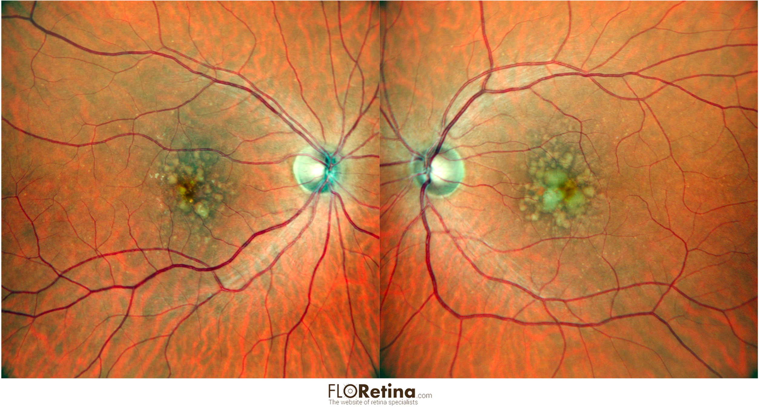
Bilateral Soft drusen
Daniela BacheriniImaging device: Color fundus photography, angle 89°, Nidek
View image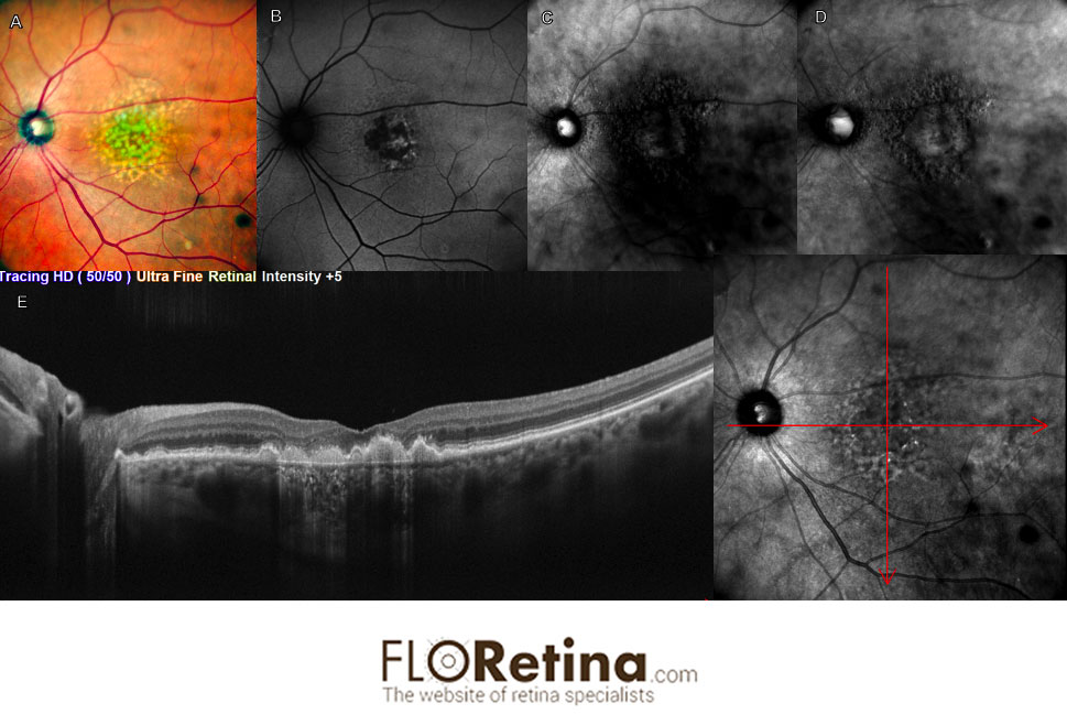
Soft drusen
Daniela BacheriniImaging device: A. Color fundus photography, angle 89°, Nidek; B. fundus autofluorescence; C. retromode DL; D. rretromode DR; E. structural OCT
View image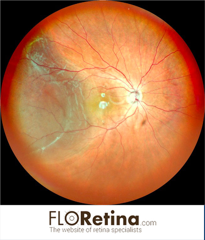
Retinal Detachment
Daniela BacheriniImaging device: Ultrawidefield color fundus photograph 163°, Nidek
View image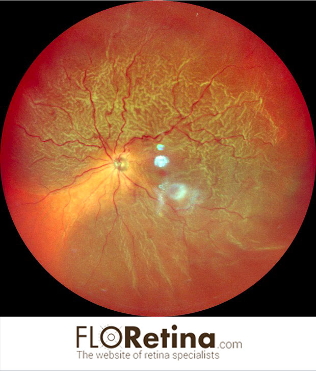
Retinal Detachment 1
Daniela BacheriniImaging device: Ultrawidefield color fundus photograph 163°, Nidek
View image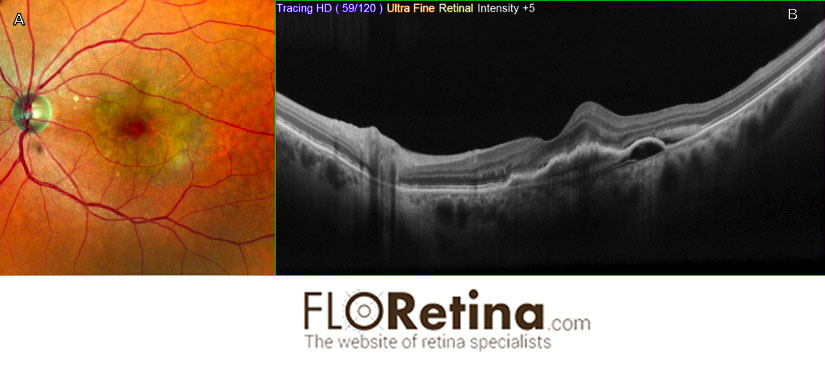
Neovascular AMD
Daniela Bacherini Francesca CipolliniImaging device: A.Color fundus photography, angle 89°, Nidek; B. Structural OCT
View image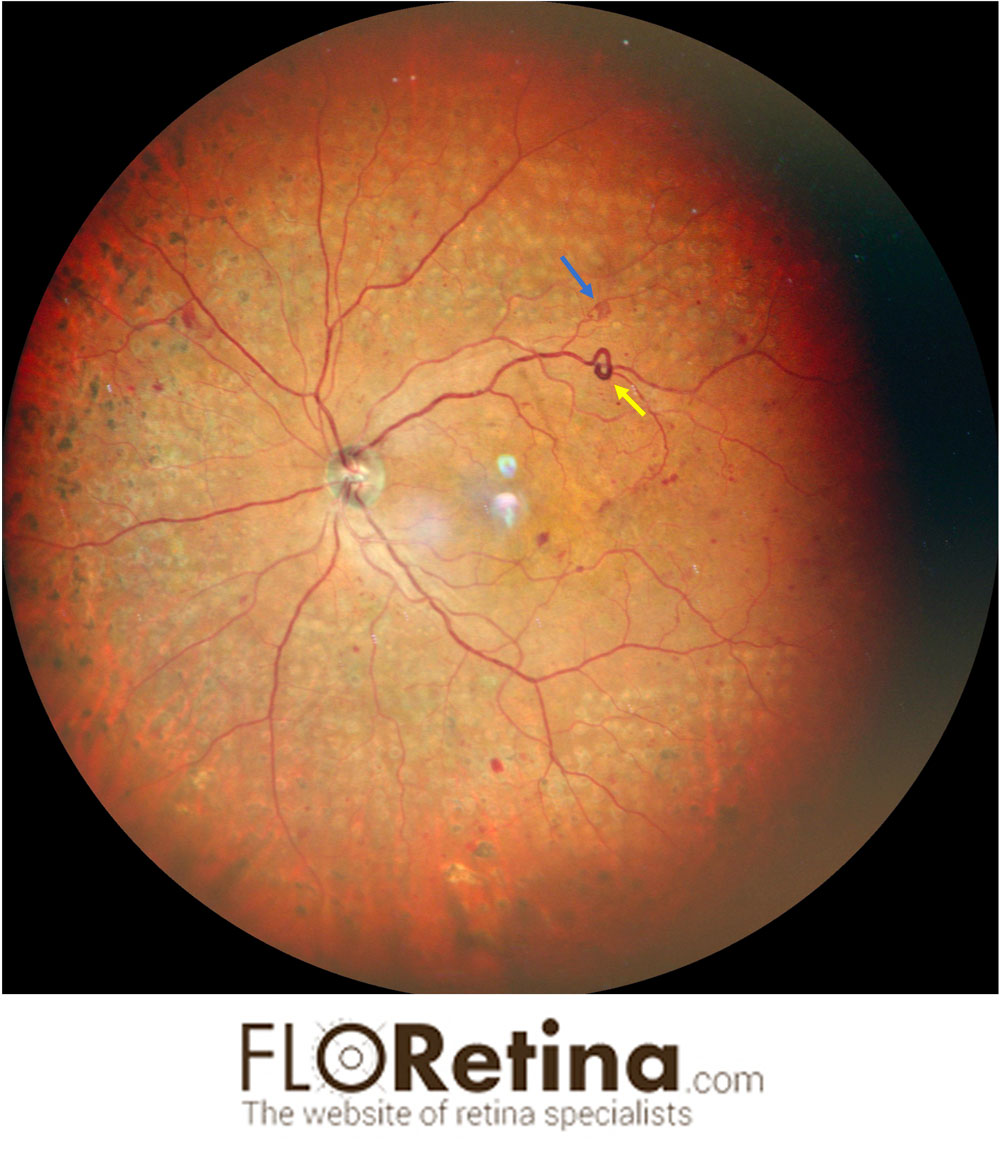
Proliferative Diabetic retinopathy
Daniela BacheriniDescription: 45 Y/O male with proliferative diabetic retinopathy treated with laser. A retinal neovessel (blue arrow), Intraretinal Microvascular Abnormality (IrMA) (yellow arrow) are evident
Imaging device: Ultrawidefield color fundus photograph 163°, Nidek
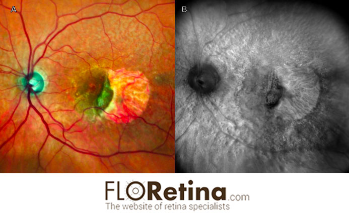
RPE tear
Daniela Bacherini Fabrizio GiansantiImaging device: A. Color fundus photography, angle 89°, Nidek, B. retromode
View image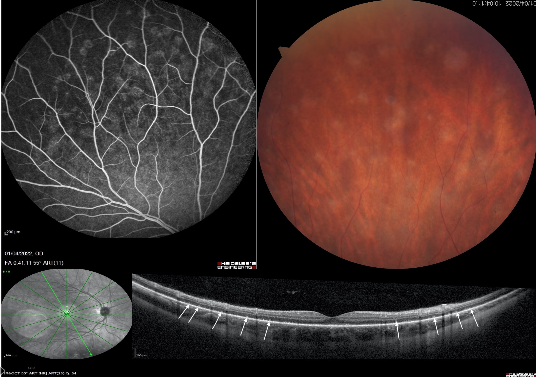
MULTIPLE EVANESCENT WHITE DOT SYNDROME.
Giovanni Romualdi Daniela BacheriniA case of 24-year-old woman with MEWDS. Structural OCT shows the loss or damage of the outer photoreceptor segments (white arrows). Hypocyanescence of the ICGA is present in the intermediate angiographic phase and most clearly detected in the late phase.
View image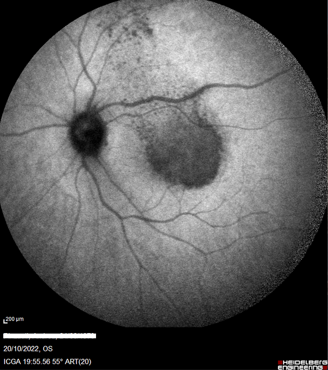
Syphilitic posterior placoid chorioretinitis
Daniela Bacherini Giovanni Romualdi Fabrizio GiansantiA case of 49 y.o. man, caucasian, with no history of drugs and systemic disease, referred to our emergency room due to a sudden para central scotoma in his left eye. Our multimodal imaging allowed us to diagnose a syphilitic posterior placoid chorioretinitis, confirmed few days later with serological exams (qualitative TPHA +).
View imageClinical case
FAST DROP IN VISUAL ACUITY IN A 47 YEARS OLD MALE: SLOW DOWN WITH THE YELLOW!
Stefano Mercuri Daniela Bacherini Gianni Virgili Fabrizio Giansanti Acute syphilitic posterior placoid chorioretinitis (ASPPC) is a rare disease which may resemble many other retinal diseases.Multimodal imaging is important for diseases characterization, and new techniques may aid us in the understanding of their etiopathogenesis.
Accurate patient’s medical patient history is fundamental to spot the correct diagnosis as soon as possible with the purpose to avoid ocular complications and complications linked to syphilis progression.
© 2026 Copyright:
Floretina.com | P.I.
01477560138
Privacy Policy
|
Cookie Policy
|
Terms and Conditions